Introduction to the morphogenesis of the salivary glands
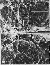
Within the human oral cavity exist three major salivary glands: the submandibular gland, the parotid gland and the sublingual gland. Each of these glands is responsible for creating a secretion which aids in food digestion and speech as well as serves as a protective immune barrier. The development of these glands follows a set pattern often seen in other organs like the lungs and pancreas where epithelial branching occurs. The salivary glands are principally induced to begin their characteristic branching morphogenesis through an interaction of the cells of the oral epithelium, which is derived from endoderm, and the underlying mesenchyme. Through various experiments, the importance of this epithelial-mesenchymal interaction has been demonstrated in conjunction with numerous proteins and other morphogenic factors. Experimental evidence reveals the importance of the extracellular matrix formed at the mesenchymal-epithelial interface and its interactions with the branching epithelium and its mesenchyme (Figure 1).
Figure 1: A high magnification view of the mesenchymal (m) -epithelial interface (e) (box 17). Box 18 is a high magnification inage of upper right hand rectangle in box 17. It is an image of the lateral surface of the lobule of the salivary gland.
The salivary glands of the mouse have provided an excellent model to examine this organ's development.
Each of the major salivary glands develops at different rates. The overall development of the parotid gland, although it is the first to begin to differentiate, takes the longest (Redman et. al, 1993). The submandibular glands are the next to appear. They begin as buds of cells in the oral epithelium and remain as such until the underlying mesoderm cues their differentiation later in development. The final of the major salivary glands to form is the sublingual gland.
The development of each of these glands follows the same general trend. For each, the endodermal epithelium first thickens into a undifferentiated cell mass which protrudes into the underlying mesoderm. As it protrudes into the mesenchyme it interacts with the basement membrane formed at the mesenchymal-epithelial interface. This basement membrane, formed from the basal lamina of the epithelium and a collagen layer secreted by the mesenchyme, contains multiple morphogenetic factors which influence branching morphogenesis. Once this branching has occurred a lumen will form in the branched stalks and lobules around the 18th day in each gland (See figure 2). This lumen allows the saliva produced by the secretory acinar cells of these glands to flow into the oral cavity and perform its stated functions. However, there is a great deal that goes on between the initial cavitation of the epithelium and the development of the lumen for active salivation.
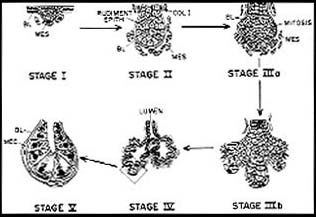
Stage I: The epithelial cell mass begins to grow into the underlying mesenchyme.
Stage II: The epithelium continues to proliferate into the mesenchyme (MES) and the basal lamina (BL), as a component of the basement membrane, separates the two layers.
Stage III:a The branching morphogenesis begins to be established as the epithelium passes through the basal lamina at the lobule ends and contacts the mesenchyme.
Stage IIIb: Collagen III is deposited along the surfaces of the cell clefts by the mesenchyme and causes the clefts to deepen. Visible contact between the epithelium and the mesenchyme at the ends of the lobules where glycosaminoglycans have degraded the basal lamina.
Stage IV: A lumen forms in the lobules and in the stalk of the salivary gland. This diagram show the apical-basal polarity of the salivary gland.
Stage V: The differentiated acinar cells which will secrete the characterizing saliva of the salivary glands. (Picture from Handbook of Physiology, 1989)
Return to Table of Contents
Return to Homepage